Arthroscopic Endoscopy: Examination and Surgery Across Key Joints
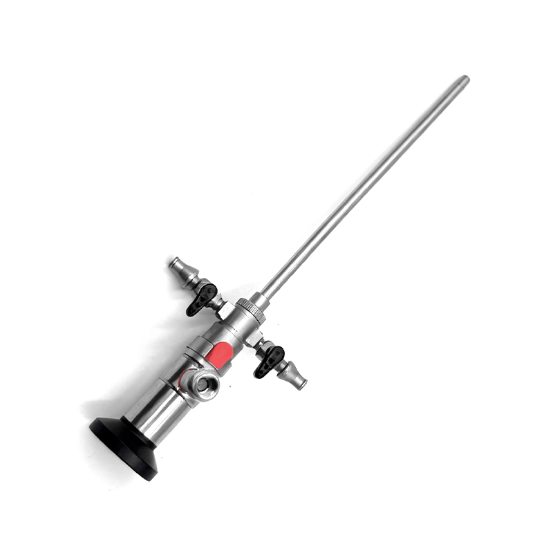
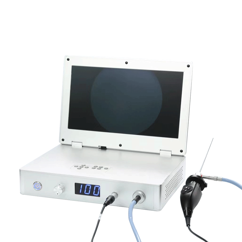
Arthroscopic endoscopy (commonly called arthroscopy) is a minimally invasive surgical technique that uses a small, fiber-optic camera (arthroscope) and specialized instruments to diagnose and treat joint disorders. Unlike open surgery, it requires only tiny incisions (5–10 mm), resulting in less pain, faster recovery, and lower infection risk. Below is a detailed overview of its application in the knee, shoulder, wrist, hip, and finger joints, including diagnostic goals and common surgical procedures.
- Knee Joint Arthroscopy
The knee is the most commonly arthroscoped joint due to its high load-bearing function and susceptibility to injuries (e.g., sports trauma, osteoarthritis).
Diagnostic Objectives
Evaluate internal structures: Menisci (cartilage cushions), anterior/posterior cruciate ligaments (ACL/PCL), medial/lateral collateral ligaments (MCL/LCL), articular cartilage, and synovium (joint lining).
Identify pathology: Meniscal tears, ligament sprains/ruptures, cartilage damage (osteochondritis dissecans), synovitis (inflammation), loose bodies (floating bone/cartilage fragments), or joint effusion (fluid buildup).
Common Surgical Procedures
Procedure Purpose Key Details
Meniscal Repair/Resection Treat torn menisci. - Repair: Sutures fix stable tears (preserves meniscal function, critical for joint protection).
- Resection (partial meniscectomy): Trim damaged tissue (for unstable/irreparable tears; avoids over-resection to prevent early arthritis).
ACL Reconstruction Restore stability after ACL rupture (common in athletes). Uses grafts (e.g., patellar tendon, hamstring tendon) to replace the torn ligament; guided by the arthroscope to ensure precise placement.
Synovectomy Relieve pain/inflammation from synovitis (e.g., rheumatoid arthritis). Remove inflamed synovial tissue to reduce joint swelling and stiffness.
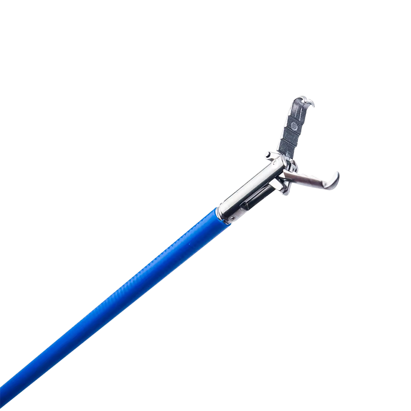
Loose Body Removal Eliminate "joint mice" (floating bone/cartilage) that cause locking or pain. The arthroscope locates fragments, which are then extracted with grasping instruments.
- Shoulder Joint Arthroscopy
The shoulder is a complex, shallow joint (ball-and-socket) prone to overuse injuries and instability. Arthroscopy is preferred here to avoid disrupting surrounding muscles (e.g., rotator cuff) in open surgery.
Diagnostic Objectives
Assess the rotator cuff (4 muscles/tendons: supraspinatus, infraspinatus, teres minor, subscapularis), glenoid labrum (fibrocartilage rim), biceps tendon, and acromioclavicular (AC) joint.
Detect tears (rotator cuff, labrum), tendinitis (e.g., supraspinatus tendinitis), impingement syndrome (acromion compressing the rotator cuff), or shoulder instability (dislocations).
Common Surgical Procedures
Rotator Cuff Repair: Reattach torn tendons to the humerus (upper arm bone) using anchors (screws) and sutures. Minimizes muscle atrophy compared to open surgery.
Labral Repair (Bankart Repair): Fixes a torn glenoid labrum (often caused by shoulder dislocations) to restore joint stability and prevent recurrent dislocations.
Subacromial Decompression: Treats impingement syndrome by removing bone spurs on the acromion or inflamed bursa (fluid-filled sac), relieving pressure on the rotator cuff.
Biceps Tenodesis: Addresses a torn or unstable biceps tendon by reattaching it to the humerus, reducing pain and "popeye" deformity.
- Wrist Joint Arthroscopy
The wrist is a delicate joint with 8 small carpal bones, making open surgery risky for nearby nerves and tendons. Arthroscopy is ideal for precise diagnosis and minimally invasive treatment.
Diagnostic Objectives
Evaluate carpal bones, intercarpal ligaments, triangular fibrocartilage complex (TFCC, a cartilage structure that stabilizes the wrist), and synovium.
Diagnose TFCC tears (common in falls on outstretched hands), carpal tunnel syndrome (median nerve compression), ligament sprains (e.g., scapholunate ligament), or arthritis (e.g., rheumatoid arthritis).
Common Surgical Procedures
TFCC Repair/Debridement: Repairs stable TFCC tears with sutures; trims damaged tissue for irreparable tears to reduce pain during wrist movement.
Carpal Tunnel Release: Relieves median nerve compression by cutting the transverse carpal ligament (the "roof" of the carpal tunnel). Arthroscopic release avoids a large palm incision, reducing scarring and recovery time.
Ligament Reconstruction: Fixes torn intercarpal ligaments (e.g., scapholunate ligament) using grafts (e.g., tendon grafts) to restore wrist stability and prevent carpal collapse.
Synovectomy: Removes inflamed synovium in arthritic wrists to reduce swelling and preserve joint function.
- Hip Joint Arthroscopy
Hip arthroscopy was once less common but has grown rapidly due to advances in instrumentation. It avoids the large incisions of open hip surgery (e.g., total hip replacement for early-stage disease).
Diagnostic Objectives
Assess the acetabulum (hip socket), femoral head (ball of the hip), labrum (acetabular rim cartilage), and hip capsule.
Detect femoroacetabular impingement (FAI, abnormal bone growth causing joint friction), labral tears (often linked to FAI), hip dysplasia (shallow acetabulum), or synovitis.
Common Surgical Procedures
FAI Correction (Cam/Pincer Resection): Removes excess bone from the femoral head (cam deformity) or acetabulum (pincer deformity) to eliminate friction and prevent further cartilage damage.
Acetabular Labral Repair: Sutures torn labral tissue to the acetabulum, restoring joint stability and reducing pain (critical for athletes or active individuals).
Hip Capsular Release: Relieves stiffness from a tight hip capsule (e.g., in athletes with overuse injuries) by cutting or stretching the capsule to improve range of motion.
Loose Body Removal: Extracts floating bone/cartilage fragments (from FAI or trauma) that cause hip clicking or locking.
- Finger Joint Arthroscopy
Finger joints (metacarpophalangeal joints, MCP; proximal interphalangeal joints, PIP) are small but essential for fine motor function. Arthroscopy is used to treat injuries from sports (e.g., basketball) or repetitive use.
Diagnostic Objectives
Evaluate joint cartilage, collateral ligaments (radial/ulnar), volar plate (stabilizes PIP joints), and synovium.
Diagnose collateral ligament tears (e.g., "skier’s thumb"—ulnar collateral ligament tear of the thumb MCP joint), PIP joint dislocations, cartilage damage, or synovitis (e.g., psoriatic arthritis).
Common Surgical Procedures
Collateral Ligament Repair/Reconstruction: Repairs torn thumb or finger ligaments with sutures; uses tendon grafts for severe tears (e.g., chronic skier’s thumb) to restore grip strength.
PIP Joint Stabilization: Fixes dislocated or unstable PIP joints by repairing the volar plate or removing loose fragments, preventing "swan-neck" or "boutonniere" deformities.
Cartilage Microfracture: Stimulates cartilage repair in small cartilage defects by creating tiny holes in the bone (releases bone marrow cells that form new cartilage).
Synovectomy: Removes inflamed synovium in arthritic finger joints to reduce swelling and preserve joint mobility (prevents permanent deformity).
Key Advantages of Arthroscopic Endoscopy (Across All Joints)
Compared to traditional open surgery, arthroscopy offers universal benefits:
Minimally Invasive: Tiny incisions reduce scarring, pain, and risk of infection.
Faster Recovery: Patients often return to daily activities within weeks (vs. months for open surgery).
Precise Visualization: The arthroscope provides high-definition, real-time images of internal joint structures, enabling accurate diagnosis and targeted treatment.
Reduced Tissue Damage: Avoids cutting through large muscles or tendons, preserving normal joint function.
Post-Operative Considerations
Recovery varies by joint and procedure but typically includes:
Immobilization: Temporary use of braces, splints, or crutches (e.g., knee/hip) to protect the joint.
Physical Therapy: Guided exercises to restore range of motion, strength, and stability (critical for long-term outcomes).
Pain Management: Over-the-counter analgesics (e.g., acetaminophen) or prescription medications (short-term) to control discomfort.
Follow-Up: Regular arthroscopic checkups to monitor healing and adjust rehabilitation plans.
 English
 عربى
عربى Español