 English
 عربى
عربى Español
Understanding the Mechanics of Unilateral Biportal Endoscopy
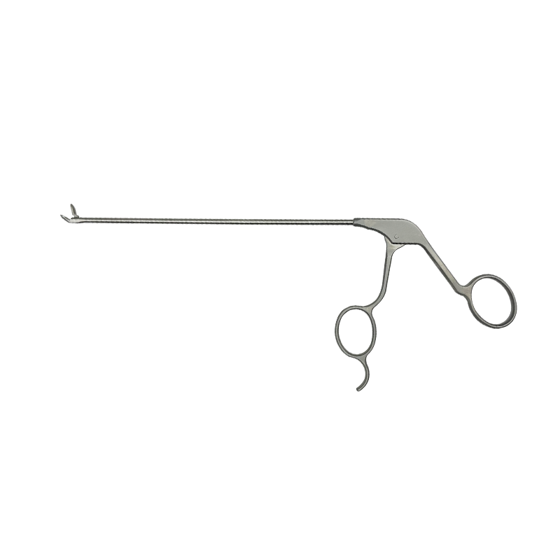
Unilateral Biportal Endoscopy, commonly referred to as UBE, represents a significant evolutionary step in spinal surgery. Unlike traditional open surgery or even some uniportal endoscopic methods, UBE utilizes two distinct channels on the same side of the spine. One portal is dedicated to the high-definition endoscope, providing a continuous, crystal-clear view of the surgical field, while the second portal serves as a dedicated working channel for surgical instruments. This separation allows for a greater range of motion and the use of conventional spinal instruments, such as high-speed drills and Kerrison rongeurs, which are often too large for single-portal systems.
The procedure is typically performed under continuous saline irrigation. This fluid pressure not only maintains a clear visual field by washing away debris and blood but also helps in controlling minor bleeding from epidural veins. By using a "floating" technique where the instruments and scope move freely within the saline-filled space, surgeons can perform delicate decompressions of the spinal canal with a level of precision that minimizes trauma to the surrounding muscle and ligamentous structures.
Clinical Indications and Patient Selection
While UBE is a versatile technique, it is particularly effective for treating degenerative conditions of the lumbar and cervical spine. Surgeons often recommend this approach for patients who have not responded to conservative treatments like physical therapy or injections. The dual-portal approach is especially advantageous in cases where extensive bony decompression is required, as it mimics the ergonomics of open surgery while maintaining a minimally invasive footprint.
Commonly Treated Conditions
- Lumbar Spinal Stenosis: Providing bilateral decompression through a unilateral approach.
- Herniated Nucleus Pulposus: Efficient removal of disc fragments causing nerve root compression.
- Foraminal Stenosis: Clearing the exit path of the nerve root to alleviate radiculopathy.
- Failed Back Surgery Syndrome: Targeted revision in patients with previous scarring.
Comparative Advantages of the UBE Technique
The adoption of UBE has grown rapidly because it bridges the gap between microscopic surgery and ultra-minimally invasive endoscopy. One of the primary benefits is the reduction in "dead space" and muscle stripping. In traditional surgery, large incisions are required to retract muscles, leading to postoperative pain and longer recovery times. UBE avoids this by entering through two small punctures, preserving the midline structures and the contralateral side of the spine.
| Feature | Traditional Open Surgery | UBE Endoscopy |
| Incision Size | 5cm - 10cm | Two 0.5cm - 1cm spots |
| Blood Loss | Moderate to High | Minimal (Saline control) |
| Hospital Stay | 3 - 5 Days | Same Day or 1 Day |
| Visualization | Microscope (External) | Endoscope (Internal/HD) |
The Surgical Workflow and Recovery Path
The UBE procedure begins with the precise placement of the two portals under fluoroscopic (X-ray) guidance. Once the triangulation of the scope and the working tool is established, the surgeon performs the laminotomy or flavectomy to access the spinal canal. Because the visual field is magnified significantly on a monitor, the surgeon can identify neural structures with extreme clarity, reducing the risk of accidental dural tears or nerve injury.
Post-surgical recovery is remarkably fast. Most patients report an immediate reduction in leg pain (radiculopathy) following the decompression. Because the paraspinal muscles are dilated rather than cut, patients are typically encouraged to walk within hours of the procedure. This rapid mobilization significantly lowers the risk of complications such as deep vein thrombosis (DVT) and allows for a quicker return to daily activities and work.
Future Perspectives and Limitations
As with any advanced surgical technique, UBE has a steep learning curve. It requires the surgeon to master "triangulation," which is the ability to coordinate the camera in one hand and the instrument in the other while looking at a 2D screen. However, as training programs become more standardized and technology improves—including the integration of 4K imaging and specialized cautery tools—UBE is expected to become a gold standard for spinal decompression.
It is important to note that UBE may not be suitable for every patient. Those with severe spinal instability requiring complex fusion, or those with significant scoliosis, may still require traditional or robotic-assisted open procedures. A thorough consultation with a spine specialist is essential to determine if the biportal endoscopic approach aligns with the patient’s specific anatomical needs and long-term health goals.


")