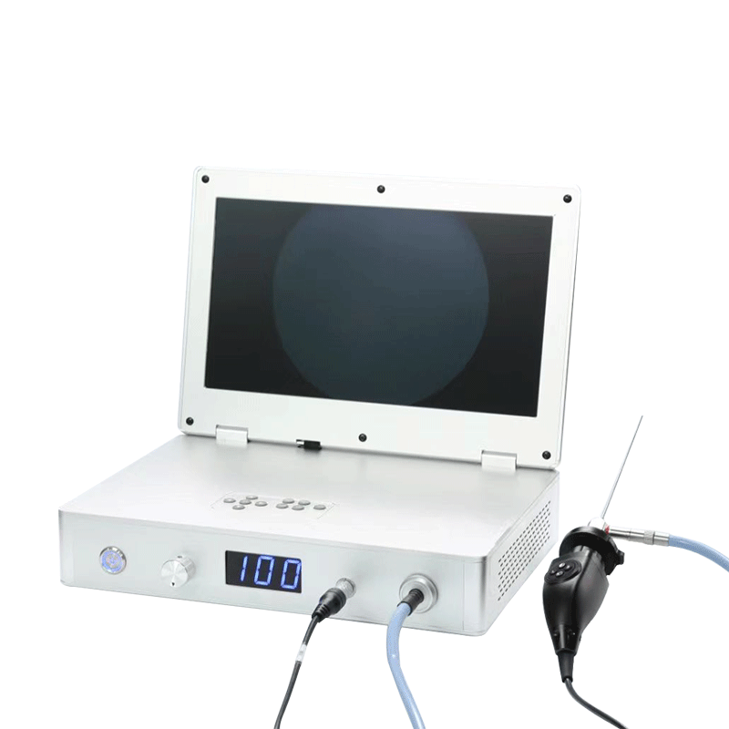
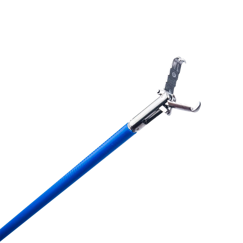
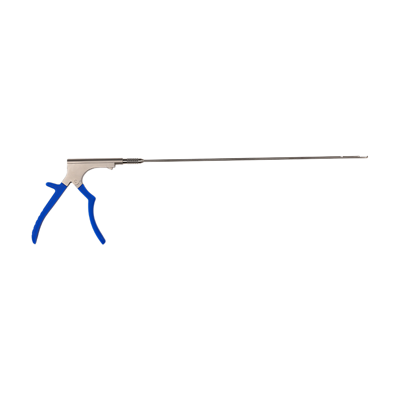
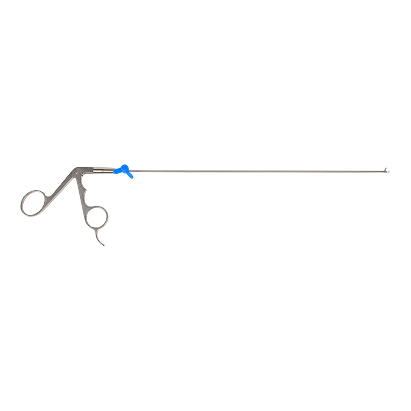
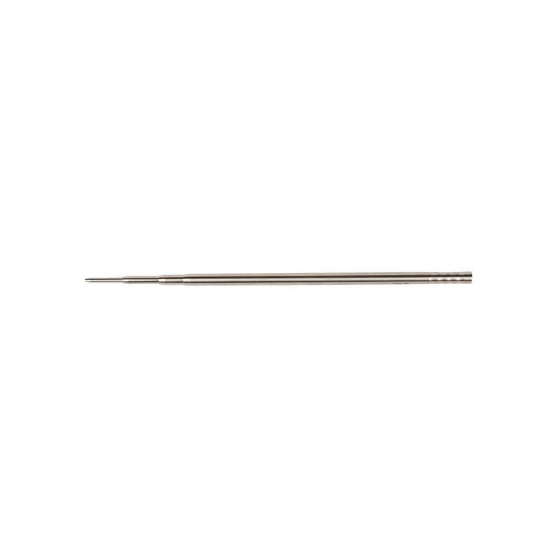
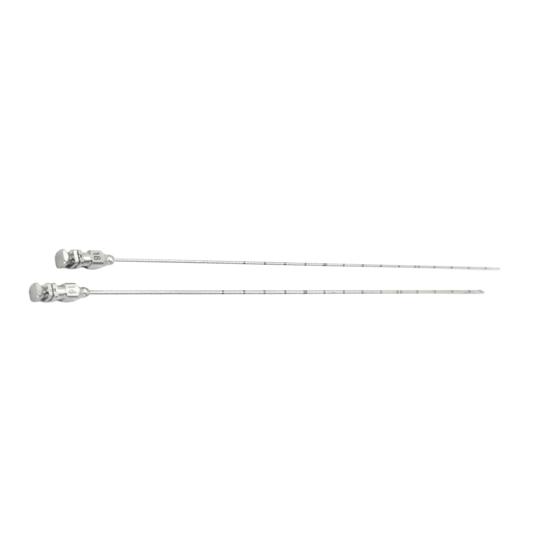
The field of spinal surgery has witnessed remarkable advancements, with a growing emphasis on minimally invasive techniques designed to reduce patient morbidity, accelerate recovery, and improve surgical outcomes. Among these innovations, the Unilateral Biportal Endoscopic (UBE) technique has emerged as a significant development, offering a versatile and effective approach to treating a wide range of spinal pathologies.
What is the UBE Technique?
The Unilateral Biportal Endoscopic (UBE) technique is a minimally invasive spinal surgical method that utilizes two separate portals – typically one for the endoscope (visualization) and another for surgical instruments (working channel). While the portals are distinct, they are positioned on the same side (unilaterally) of the patient's body, allowing the surgeon to operate with enhanced maneuverability and a clear, magnified view of the surgical field. This contrasts with traditional open surgery, which requires larger incisions, and even some other endoscopic techniques that might use a single port or multiple ports on different sides.
Key Principles and Advantages
The UBE technique leverages several fundamental principles that contribute to its growing popularity:
-
Separate Portals for Enhanced Dexterity: The distinct camera and working portals provide surgeons with greater freedom of movement and triangulation of instruments. This allows for more precise manipulation of tissues, decompression of neural structures, and placement of implants compared to single-port systems.
-
Optimal Visualization: The endoscope delivers a high-definition, magnified view of the surgical anatomy, including neural elements, bone, and soft tissues. Irrigation through the scope continuously clears the field, ensuring excellent visibility throughout the procedure.
-
Minimally Invasive Nature: By utilizing small incisions (typically 5-8 mm each), the UBE technique minimizes muscle disruption, blood loss, and postoperative pain. This translates to faster recovery times, shorter hospital stays, and a quicker return to daily activities for patients.
-
Versatility: UBE is highly adaptable and can be applied to various spinal levels (cervical, thoracic, and lumbar) and a multitude of conditions.
-
Reduced Risk of Complications: Compared to open surgery, the smaller incisions and enhanced visualization often lead to a lower risk of infection, nerve damage, and other common surgical complications.
-
Cost-Effectiveness: While initial setup may involve specific equipment, the reduced hospital stay and quicker recovery can contribute to overall cost savings for the healthcare system and the patient.
Indications for UBE
The UBE technique has proven effective in addressing a broad spectrum of spinal conditions, including but not limited to:
-
Lumbar Disc Herniation: Decompression of the nerve root impinged by a herniated disc.
-
Lumbar Spinal Stenosis: Decompression of the spinal canal to relieve pressure on the spinal cord and nerve roots. This can include central, lateral recess, and foraminal stenosis.
-
Spondylolisthesis (low-grade): Decompression and potentially fusion (if instrumentation is used).
-
Foraminal Stenosis: Widening of the neural foramen to relieve nerve root compression.
-
Cervical Radiculopathy/Myelopathy: Decompression in the cervical spine, though this application is more technically demanding.
-
Thoracic Disc Herniation: A less common but viable indication.
-
Synovial Cysts: Resection of cysts causing neural compression.
-
Spinal Tumors (select cases): Biopsy or removal of certain benign tumors.
The Surgical Procedure (General Steps)
While specific steps may vary depending on the pathology and surgeon's preference, a typical UBE procedure involves:
-
Patient Positioning: The patient is positioned prone on a radiolucent table, allowing for fluoroscopic guidance.
-
Incision and Portal Placement: Two small incisions are made on the unilateral side, typically 1-2 cm apart. One incision serves as the entry point for the endoscope, and the other for surgical instruments.
-
Working Space Creation: Saline irrigation is continuously infused to distend the working space and clear the field of blood and debris, creating a clear "aquatic" surgical environment.
-
Target Visualization and Decompression: Under direct endoscopic visualization, the surgeon uses various specialized instruments (e.g., kerrisons, burrs, dissectors, graspers) to perform the necessary decompression, disc removal, or other surgical maneuvers.
-
Hemostasis: Careful hemostasis is maintained throughout the procedure.
-
Closure: Once the surgical objectives are achieved, the instruments and endoscope are removed, and the small incisions are closed with sutures or steristrips.
Postoperative Care and Recovery
Due to its minimally invasive nature, UBE typically allows for a relatively swift recovery:
-
Pain Management: Patients usually experience less postoperative pain compared to open surgery and can often be managed with oral analgesics.
-
Early Mobilization: Patients are encouraged to ambulate shortly after surgery.
-
Hospital Stay: Often a same-day discharge or an overnight stay.
-
Rehabilitation: A structured physical therapy program may be recommended to strengthen core muscles and restore full function.
-
Return to Activity: Most patients can return to light activities within a few days to a week and more strenuous activities within 4-6 weeks.
Future Directions and Conclusion
The Unilateral Biportal Endoscopic technique represents a significant leap forward in minimally invasive spinal surgery. Ongoing research and technological advancements continue to refine the instruments and techniques, expanding its applications and improving outcomes. As surgeons gain more experience and training in UBE, it is poised to become an increasingly standard and preferred approach for a wide array of spinal conditions, ultimately benefiting patients through less invasive procedures, reduced recovery times, and improved quality of life.


 English
 عربى
عربى Español