 English
 عربى
عربى Español
A urology resectoscope is an essential instrument in the field of urology, used for a variety of endoscopic surgical procedures within the urinary tract. This specialized tool allows surgeons to operate with precision and minimal invasiveness, offering significant benefits to patients compared to traditional open surgery. The resectoscope combines the functions of a cystoscope and a surgical tool, making it a versatile and indispensable device.
The Design and Components
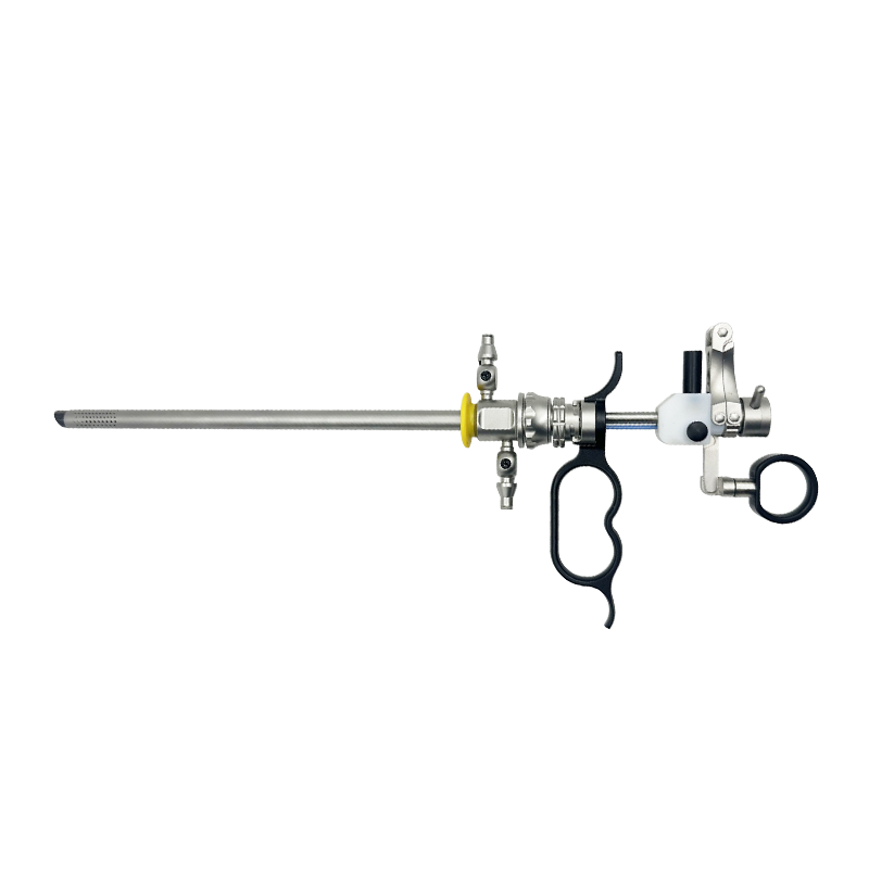
At its core, the resectoscope is a long, thin instrument with a camera (telescope) and a light source at its tip. This allows the surgeon to visualize the internal structures of the bladder, prostate, or urethra on a monitor. The resectoscope also includes a working channel through which various surgical loops and electrodes can be passed. These loops, often powered by an electrosurgical generator, are used to cut, vaporize, or coagulate tissue.
Key components of a standard resectoscope include:
-
Telescope: Provides a magnified, clear view of the surgical site.
-
Sheath: The outer tube of the instrument that allows for the passage of fluid and instruments.
-
Working Element: The part of the resectoscope that holds the surgical loop and allows the surgeon to manipulate it.
-
Electrosurgical Loop: The cutting or coagulating wire used to perform the surgery.
Common Applications
The primary use of a urology resectoscope is in transurethral surgery, meaning the procedure is performed by inserting the instrument through the urethra without making an external incision. This approach is fundamental to modern urological care.
Some of the most common applications include:
Transurethral Resection of the Prostate (TURP)
This is perhaps the most well-known application of the resectoscope. TURP is the gold standard for treating benign prostatic hyperplasia (BPH), a condition where the prostate gland enlarges and obstructs urine flow. The surgeon uses the resectoscope's cutting loop to carefully remove excess prostatic tissue, creating a wider channel for urination.
Bladder Tumor Resection
The resectoscope is also used for the diagnosis and treatment of bladder tumors, in a procedure known as Transurethral Resection of Bladder Tumor (TURBT). The surgeon can resect the tumor and surrounding tissue while also obtaining tissue samples for pathology, which is crucial for determining the type and stage of cancer.
Urethral Stricture Management
In cases of urethral stricture—a narrowing of the urethra—the resectoscope can be used to incise the scar tissue causing the blockage. This procedure, known as a direct visual internal urethrotomy (DVIU), helps to restore normal urine flow.
Advances in Resectoscope Technology
Over the years, the urology resectoscope has evolved with technological advancements. Modern resectoscopes may feature:
-
Bipolar Energy: Traditional resectoscopes use monopolar energy, which requires a grounding pad and can increase the risk of complications. Bipolar resectoscopes use two electrodes within the loop, which allows for surgery to be performed in saline irrigation fluid. This significantly reduces the risk of complications like TUR syndrome.
-
Continuous Flow Systems: These systems ensure a constant flow of irrigation fluid, which helps to maintain a clear visual field and wash away blood and tissue debris.
-
High-Definition Cameras: Advances in optics and camera technology provide surgeons with an even clearer, more detailed view of the anatomy, enhancing precision and safety.
In conclusion, the urology resectoscope remains a cornerstone of minimally invasive urological surgery. Its ability to provide both visualization and surgical capabilities through a single, non-incisional approach has revolutionized the treatment of numerous conditions, improving patient outcomes and recovery times.


")